
Achilles Tendinopathy
What is the Achilles tendon?
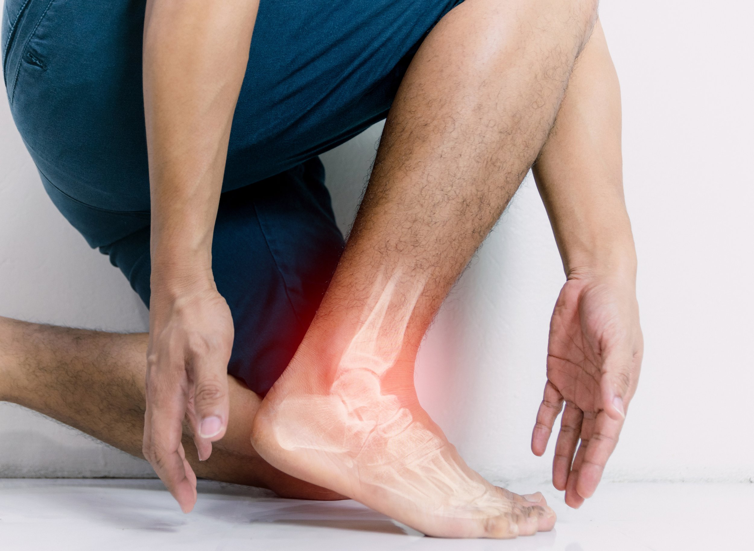
Your Achilles tendon is an important part of your leg. It is found just behind and above your heel. It joins your heel bone (calcaneum) to your calf muscles. The function of your Achilles tendon is to help in bending your foot downwards at the ankle: this movement is called plantar flexion.
What is Achilles tendinopathy and what causes it?
Two types of Achilles tendinopathy are commonly described:
1. Non–insertional Achilles tendinopathy, where the mid portion (2-6cm from the insertion to the calcaneous/heel bone) is affected.
2. Insertional Achilles tendinopathy, tendinopathy up to 2cm from the Achilles tendon insertion to the calcaneous/heel bone is affected.
This leaflet describes both conditions and their treatment options.
Achilles tendinopathy is a condition that causes pain, swelling and stiffness of the Achilles tendon. It is thought to be caused by repeated tiny injuries (known as micro-trauma) to the Achilles tendon. After each injury, the tendon does not heal completely, as should normally happen. This means that over time, damage to the Achilles tendon builds up and Achilles tendinopathy can develop.
There are a number of things that may lead to these repeated tiny injuries to the Achilles tendon, for example:
∙ Overuse of the Achilles tendon. This can be a problem for people who run regularly. Achilles tendinopathy can also be a problem for dancers and for people who play a lot of tennis or other sports that involve jumping.
∙ Being overweight.
∙ Training or exercising wearing inappropriate footwear.
∙ Having poor training or exercising techniques - for example, a poor running technique.
∙ Making a change to your training programme - for example, rapidly increasing the intensity of your training and how often you train.
∙ Training or exercising on hard or sloped surfaces.
Achilles tendinopathy is also more common in people who have certain types of arthritis, such as ankylosing spondylitis or psoriatic arthritis. It is also thought that your genetic 'makeup' (characteristics inherited from your parents), may play a part for some people who develop Achilles tendinopathy. It is also more common in people who have high blood pressure, high cholesterol or diabetes
People who are taking medicines from a group called fluoroquinolones (for example, the antibiotics ciprofloxacin and ofloxacin), also have an increased risk of developing Achilles tendinopathy.
Achilles tendinopathy used to be known as Achilles tendonitis. In general, 'itis' usually refers to inflammation, so tendonitis would mean inflammation of a tendon, however Achilles tendinopathy is now thought to be a better term to use because it is thought that there is little or no inflammation that causes the problem.
If the Achilles tendon is torn, this is called an Achilles tendon rupture. How common is Achilles tendinopathy?
About 6 in 100 inactive people develop Achilles tendinopathy at some point in their lifetime. However the chance of it developing is higher in athletes or those who train regularly or do a lot of exercise. It can be a particular problem for some runners. It used to be thought that it is more common in men than in women but this may not be true.
What are the symptoms of Achilles tendinopathy?
The main symptoms include pain and stiffness around the affected Achilles tendon. Pain and stiffness tend to develop gradually and are usually worse when you first wake up in the morning. Severe pain that comes on suddenly and difficulty walking can be symptoms of Achilles tendon tear (rupture).
Some people have pain during exercise but, in general, pain is worse after exercise. Runners may notice pain at the beginning of their run, which then tends to ease and become more bearable, followed by an increase in pain when they have stopped running. Pain due to Achilles tendinopathy may actually prevent you from being able to carry out your usual everyday activities such as walking to the shops, etc. You may notice that you have pain when you touch the area around your Achilles tendon. There may also be some swelling around this area.
Do I need any investigations?
Your doctor will usually diagnose Achilles tendinopathy because of your typical symptoms and from examining your Achilles tendon. They may feel for swelling or tenderness of the tendon. They may also ask you to do some exercises to put some stress on your Achilles tendon. For example, they may ask you to stand on the affected leg and raise your heel off the ground. For most people with Achilles tendinopathy this movement brings on (reproduces) their pain. If this does not bring on your pain, your doctor may ask you to hop on that foot, either on the spot or in a forwards direction. Your doctor may also do some other tests to make sure that there are no signs that you have torn (ruptured) your Achilles tendon. For example, squeezing your calf muscles and looking at how your foot moves.
X-rays or other tests are not usually needed to diagnose Achilles tendinopathy however an ultrasound scan or an MRI scan may sometimes be suggested by your specialist if the diagnosis is not clear or to aid surgical planning.
What is the initial treatment for Achilles tendinopathy?
There are a number of treatments that may help both insertional and non insertional Achilles tendinopathy. The treatments below are usually suggested first. They are all considered as conservative treatments (treatments that do not involve surgery).
Rest
Rest and time off from sporting activities are important if you have Achilles tendinopathy. At first, you should stop any high-impact activities or sports (such as running). As pain improves, you can restart exercise as your pain allows. It is thought that complete rest, if it is prolonged, can actually be worse for the injury. Talk to your doctor or physiotherapist about when you should start exercising again.
Pain killers
Pain killers such as paracetamol or ibuprofen may help to relieve pain. Ibuprofen is from a group of medicines called non-steroidal anti-inflammatory drugs (NSAIDs) however you should not use ibuprofen or other NSAIDs for more than 7-14 days if you have Achilles tendinopathy. This is because they may possibly reduce the ability of the tendon to heal in the long term. They may also cause symptoms of Achilles tendinopathy to be masked, or covered up, which again may delay healing.
Note: Side-effects sometimes occur with anti-inflammatory painkillers. Stomach pain and bleeding from the stomach is the most serious. Some people with asthma, high blood pressure, kidney failure and heart failure may not be able to take anti-inflammatory painkillers, so check with your doctor or pharmacist before taking them if you are unsure if they are suitable for you.
Ice packs
Ice treatment may be useful for pain control and may help to reduce swelling in the early stages of Achilles tendinopathy. An ice pack should be applied for 10- 30 minutes: less than 10 minutes has little effect and more than 30 minutes may damage the skin. Make an ice pack by wrapping ice cubes in a plastic bag or towel. Do not put ice directly next to skin, as it may cause ice burn. A bag of frozen peas is an alternative. Gently press the ice pack on to the injured part.
The cold from the ice is thought to reduce blood flow to the damaged tendon, this may reduce pain. Do not leave ice on while asleep.
Achilles tendon exercises
Some special exercises to help to stretch and strengthen your Achilles tendon have been proven to be helpful. You should aim to do these every day. Such exercises may help with pain control and stiffness. A physiotherapist may be
able to help you with these exercises as needed. They may also use other treatments such as ultrasound and massage to help relieve symptoms and promote healing of your Achilles tendon.
The following exercises can be used to help treat Achilles tendinopathy:
∙ Stand about 40 cm away from a wall and put both hands on the wall at shoulder height, feet slightly apart, with one foot in front of the other. Bend your front knee but keep your back knee straight and lean in towards the wall to stretch. You should feel your calf muscle tighten. Keep this position for several seconds, and then relax. Do this about 10 times and then switch to the other leg. Now repeat the same exercise for both legs but, this time, bring your back foot forward slightly so that your back knee is also slightly bent. Lean against the wall as before, keep the position, relax and then repeat 10 times before switching to the other leg. Repeat this routine twice a day.
∙ Stand on both feet. Use your unaffected leg to rise up on to tiptoes. Transfer your weight across to your affected leg and lower yourself down. Repeat and aim for three sets of 15 repetitions twice each day. The standing on tiptoe exercise should be performed with your knees straight and with your knees bent.
∙ Stand on the bottom step of some stairs (facing upstairs) with your legs slightly apart and with your heels just off the end of the step. Hold the stair rail for support. Rise up on tiptoes, then, standing on the affected leg, lower your heel, keeping your knee straight. Put your good leg down before lifting your heel. Repeat and aim for three sets of 15 repetitions twice each day. Repeat but this time keeping the knee of the affected leg slightly bent. Try to do these exercises twice a day.
∙ Keep a towel (or a long piece of elastic) by the side of your bed. First thing in the morning, before you get out of bed, loop the towel around the ball of one of your feet then pull the towel towards you, keeping your knee straight. Hold the pull for about 30 seconds. This exercise will pull your toes and the upper part of your foot towards you. Repeat this exercise three times for each foot.
∙ Sit on a chair with your knees bent at right angles and your feet and heels flat on the floor. Lift your foot upwards, keeping your toes on the floor. Hold the position for a few seconds and then relax. Repeat about 10 times. Try to do this exercise five to six times a day.
Orthotics
An orthotics specialist may suggest changing your footwear or putting special inserts in your shoes, such as inserts to lift your heel. This may help to reduce pain and symptoms.
What happens if initial treatment fails?
For most people, the symptoms of Achilles tendinopathy usually clear within 3-6 months of starting conservative treatment, as described above. In general, the earlier the problem is recognised and treatment started, the better the outcome.
If your symptoms have not improved after 3-6 months your specialist may recommend the following treatment options.
Specialist treatments that are not surgery
∙ Steroid injections: Injection of a steroid medicine is a common treatment for some tendon injuries. However, the use of steroid injections for the treatment of Achilles tendinopathy is controversial in the UK.
∙ Extracorporeal shock-wave therapy (see separate leaflet): During this treatment, special sound waves are passed through your skin to your Achilles tendon. Side-effects can include reddening of your skin and an ache in your calf after treatment. This procedure is generally safe but there is a small risk of tearing (rupturing) the Achilles tendon with this treatment, particularly in older people. At the moment, it is uncertain how effective this treatment is and how well it works. The National Institute for Health and Care Excellence
(NICE) therefore recommends that specialists discuss this treatment fully with anyone before it is started.
Are there any complications of Achilles tendinopathy?
There is a risk of tearing (rupturing) the Achilles tendon if you have Achilles tendinopathy. This is because the tendon is damaged and weaker than usual however this risk is usually quite low. Severe pain around the Achilles tendon that develops suddenly may be a sign of tendon rupture. See a doctor urgently if you think that you may have ruptured your Achilles tendon.
Surgery for non-insertional (mid portion) Achilles tendinopathy
Those who have persisting pain due to Achilles tendinopathy that has not responded to other treatments may have surgery to treat the condition. Most people have a good result from surgery and their pain is relieved. Surgery involves either of the following:
• Removing nodules or adhesions (parts of the fibres of the tendon that have stuck together) and have developed within the damaged tendon (debridement).
• Making lengthways or multiple small cuts in the tendon to help to stimulate and encourage tendon healing.
Complications from surgery are not common, but if they do occur, can include problems with wound healing/infection and tendon rupture.
The goal of surgery is to eliminate pain and improve a patient’s mobility. Surgery is commonly performed under general anaesthetic and in combination with a nerve block which helps reduce post-operative pain. Regional anaesthetics are also possible and may be discussed with you by your anaesthetist.
Insertional tendinopathy
This is a degeneration of the fibers of the Achilles tendon directly at its insertion into the heel bone. It may be associated with inflammation of a bursa in the same area. The treatment options initially are similar to those detailed previously, but may progress to surgery that involves excision of the so called “pump bump” at the back or side of the heel.
What are the symptoms?
Most patients report the gradual onset of pain and swelling at the Achilles tendon insertion into the back of the heel bone without specific injury. At first, the pain is noted after activity alone, but becomes more constant over time. The pain is made worse by jumping or running and especially with sports requiring short bursts of these activities. There is tenderness directly over the back of the heel bone and often there is a bone prominence at this area.
How is insertional Achilles tendinopathy diagnosed?
This remains primarily a clinical diagnosis. X-rays may show calcification deposits within the tendon at its insertion into the heel. X-rays may also demonstrate the Haglund's deformity.
Magnetic resonance imaging (MRI) remains the imaging option of choice because it can determine the extent of tendon degeneration as well as other factors such as bursitis, which may contribute to pain at the base of the heel.
∙ Non-surgical treatment options: Conservative non-surgical treatment as detailed in the previous section (non-insertional tendinopathy) remains effective in the majority of patients with liberal use of nonsteroidal anti inflammatory drugs, heel lifts, stretching and shoes that do not provide pressure over this area. If symptoms persist, appropriate orthotics and physical therapy may be of benefit. If this fails, then application of a cast or brace with gradual return to activity may be indicated.
∙ Surgical treatment options: Surgical treatment is indicated if there is failure of several months of nonsurgical treatment. Surgery removes the degenerative portions of the tendon, any bone which is irritating the tendon and any inflamed bursa tissue. If the tendon is short, then lengthening may also be necessary. The tendon attachment to the heel bone may need to be strengthened with sutures that attach directly into the bone.
How long is recovery after surgery?
After surgery, a plaster cast is worn for two weeks often in a toe-down position to allow wound healing. Once the wound begins to heal, weight bearing in a cast or boot in a toe-down position commences. Physical therapy may be started from six weeks. Return to athletic activities usually occurs from 12 weeks. Post op rehabilitation/physiotherapy will often be bespoke depending on the amount of detachment of the tendon at the time of surgery.
Some patients may require one to two years to recover fully following both surgical and non-surgical treatment. Good to excellent results after surgery are about 75-85 percent.
Potential complications
Because of the poor blood supply to the skin and tendon in this area, the greatest risk following surgery is that of wound complications, infection and tendon detachment.
Before the operation (pre-operatively)
You will see a pre-admission nurse to check you are medically well enough for surgery. It is important to mention any medicines that you are taking, either prescribed or non-prescribed, including over the counter medications, herbal remedies or aspirin, warfarin, hormone replacement therapy (HRT), the contraceptive pill or medication for high blood pressure.
Prior to admission for your surgery there are a number of issues that need to be considered, for example, can someone help you carry out basic everyday tasks such as preparing food and food shopping? If you have stairs, how will you get up and down them? Do you have sturdy hand rails? If your toilet is downstairs, would it be easier to have your bed downstairs until you have recovered and are able to negotiate the stairs safely?
The pre-admission nurse will discuss these with you and if they have any concerns about your coping at home after your operation, they may refer you to a physiotherapist and/or an occupational therapist. The therapist will telephone you to discuss your needs and it may be necessary to attend for a more in-depth assessment. This will ensure that we plan for your discharge home safely and shorten your stay in hospital.
What to bring with you
Please ensure that you have a flat sturdy shoe to wear on the non-operated foot following surgery. If you use a walking stick or crutches, please ensure you bring these with you.
After both types of Achilles tendon surgery
You will usually stay in hospital for approximately one to two days after your operation. A physiotherapist will see you on the ward and teach you how to walk using a walking aid. If you have to use stairs at home you will be taught the safest way to do this.
Treatment is individual for each patient. However, it is normal, following Achilles tendon surgery, for you to have a plaster cast in place following your operation. Your foot may be placed in a plantar flexed (toes pointing down) position. You will not be allowed to bear weight upon the operated leg (initially)
Outpatient review
You will be given an appointment to attend the Outpatients’ Department two weeks following your procedure. The sutures will be removed and your wound site will be inspected. A new plaster or boot will then be reapplied.
Your plaster cast may be changed every two weeks until your foot is in a neutral position. You may be non-weight bearing (this means NO weight is to be put through the affected limb) until instructed. When the foot has reached a neutral position you may put your full weight through the operated limb while walking. This is achieved about 4-6 weeks after the operation. You will come out of the plaster or boot completely six to twelve weeks after the operation (depending on the operation you have had).
Prolonged physiotherapy is required following your operation. You may also need to have an insole for your shoe and this will be arranged with the orthotist should it be necessary.
Things to look out for
• Swelling: You should expect some swelling after your operation. If swelling persists or worsens and you are concerned, seek advice from a member of the foot and ankle team (contact details below) or your GP.
• Infection: Any operation is at risk of infection. Fortunately it is not common in this type of surgery but a small number of patients do get a wound infection and these normally settle after a short course of antibiotics. In rare circumstances, the infection may be more severe and require further surgery to remove infected tissue and administer a longer course of antibiotics.
• Blood clots: Deep Vein Thrombosis (DVT) or Pulmonary Embolus (PE) are rare but can occur. Please inform the team if you have had a DVT or PE in the past or if you have a family history of clotting disorders. You will be given an anti-embolic stocking to wear on your other leg and blood thinning injections while your leg is in plaster.
• Numbness or tingling: This can occur at the surgical site(s) if fine, hair-like nerves are cut or more major nerves are stretched. This is normally temporary; however, patchy numbness or sensitised areas may be permanent. In rare circumstances, the nerves can become hypersensitive in a condition called ‘Complex Regional Pain Syndrome’. This can lead to severe pain as well as colour and temperature changes in the foot. If this happens, your consultant will discuss treatment with you.
• Wound healing: If blood supply to the area is not good, wounds may be slow to heal. If this is the case, more frequent wound dressings may be required to ensure that the wound does not become infected.
• Scarring: Any type of surgery will leave a scar. Occasionally this can cause pain and irritation. If this happens, please discuss with your consultant.
Getting back to normal
• Returning to work: If your job is mostly sitting, you may be allowed back to work four weeks following surgery, provided you can keep the leg elevated. However, if your job is more physical and involves long periods on your feet then it may take longer.
• Walking: Most people aim to be walking independently three months after the operation. However this depends on your walking tolerance before the operation and your progress following.
• Footwear: It can take several months for swelling to go down, but most people can wear normal footwear by six months.
• Driving: If you have an automatic car and undergone surgery on the left foot, you can usually drive by two weeks after your operation. Otherwise, it may take about three months to be able to drive. In order to be safe to drive, you must be able to perform an emergency stop. You must inform your insurance company regarding the type of operation that you have undergone to ensure that your cover is valid.
• Sport: Resuming sporting activity depends on your operation and will be discussed with you. Generally you can return to low impact sports approximately three to six months after your operation, but it may be as much as one year before you are fully recovered.